Back
References: Honda A et al, Isolation, characterization, and
in vitro and in vivo differentiation of putative thecal stem cells. Proc
Natl Acad Sci USA 2007;104:12389.
Summarized by: Mary Dovlatyan, Fall 2007
LAY REVIEW
Thecal cells are specialized cells that make up a layer of the ovarian
follicles. Their primary purpose is the production of a hormone that is the
precursor to estrogen and structural support which are performed by the theca
interna and theca externa cells, respectively. Originally the primary goal of
the authors was to isolate female germ line stem cells. Germ cells are those
cells that contain half the DNA necessary for reproduction. They come from
either a male or female and produce the egg and sperm of an organism. The
discovery is that the apparent stem cells were instead, somatic cells rather
than germ cells. Thus, the authors proceeded to characterize the isolated
somatic cells as well as functional studies.
The thecal stem cells were isolated from ovaries of newborn mice, 2-4 days
old ICR and green fluorescent mice. Granulosa cells that were not contaminated
with thecal cells were also studied. Characterization was addressed by electron
microscopy and gene expression analysis, which led to the supposition of theca
stem cells.
Functional studies were determined in the ability of the cells to
differentiate by using culture conditions that induce differentiation. For
example when serum was added, the round colonies spread and appeared to begin
hormone production. Additional growth factors led to the observation of specific
organelles. The isolated granulosa cells, stated above, were added to the
culture. This induced further differentiation of the theca stem cells. The newly
formed granulosa cells were identified from the added granulosa cells by added
granulosa cells from the “green” mice. This would be different from granulosa
cells that are not expected to be green. The level of androstenedione, which is
the hormone necessary for estrogen production, in the media also increased as
the thecal cells differentiate.
The final stage in the authors’ study was to perform intraovarian
transplantation of green thecal stem cells into non-green mice. The green stem
cells were identified in the ovaries by fluorescence microscopy. Finally the
host mice were given equine chorionic gonadotropin to produce follicular
development. When the ovaries were removed and observed the green fluorescent
cells proliferated and surrounded the fully developed follicles.
I found that this article informative and well written. More importantly, the
authors clearly outlined their thought processes. They began with the studies
with the intent to do something completely different and when they hit the
obstacle of the cells being somatic cells rather than germ cells. The authors
instead, used this obstacle as an opportunity to study something new. Also they
raised questions throughout the article and then performed experiments to
address their questions. For example if these were not germ cells what kind of
stem cells were they? They used microscopy and gene expression profiling. One
question is when they were applying the cells to different growth factors did
true differentiation occur or was it an artifact of the media? I believe that
these cells were actually stem cells because they were able to culture multiple
passages and showed success in their transplantation. I believe that the
addition of different growth factors created a microenvironment that facilitated
differentiation. For example in the last stage they showed that granulosa cells
were needed for final differentiation. In ovaries, granulosa cells, which are
located next to the thecal cells may therefore be cellular component of the
microenvironment for the differentiation of natural thecal cells. It would be
wonderful to see future studies in animal models for translational studies with
thecal stem cells. Also they could use these experiments to further study the
effect of the microenvironment on stem cells and perhaps apply the findings to
other types of adult stem cells.
SCIENTIFIC REVIEW
Thecal cells are specialized cells that make up a layer of the ovarian
follicles. Their primary purpose is hormone production and structural support
which are performed by the theca interna and theca externa cells respectively.
Originally the primary goal of the authors was to isolate female germ line stem
cells. However, instead of germ stem cells, the cells were somatic. The
authors proceeded to characterize the isolated somatic cells and also performed
functional in vitro and in vivo studies.
To obtain the thecal stem cells the authors initially collected ovaries from
newborn mice that were 2-4 days old for cell culture. The in vivo studies were
done with ICR and “green” fluorescent mice. When culturing these cells they were
able to perform four to five passages before the cells stopped proliferating.
They also cultured granulosa cells and eliminated contamination with thecal
cells by molecular analyses. During the initial isolation of the thecal cells, a
serum-free medium was used containing growth factors. The floating cells were
then passaged to a second culture. The second culture resulted in small round
colonies and stained similar to other stem cells. The cultured cells were also
treated with BrdU. The small cell colonies that stained positive for BrdU,
which indicated mitotic activity did not stain for MVH (a germ cell marker).
The results indicated that the cells within the colonies were somatic cells
rather than germ cells. In the next step, the authors identified the somatic
cells as interstitial, based on electron microscopy. These cells were also
distinguishable from granulosa cells by alkaline phosphatase reactions. Finally,
gene expression analysis determined the expressions of theca cell markers, Ptch1
and Gli3, but not markers of granulosa cells. In total, the results suggested
the isolation of thecal stem cells.
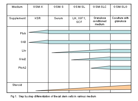
The next step performed in vitro differentiation of the theca stem
cells from serum-free media. This was done by replating the cultured cells in
five different conditions. Each condition contained serum and different growth
factors. Each condition resulted in more matured cells, indicating variations
of cell maturity. For example, when serum was added the original round colonies
spread and appeared to begin steroidogenesis. As more growth factors were added,
the cells showed higher levels of differentiation, which was apparent by the
observation of organelles specific for steroidogenesis. As stated earlier
granulosa cells were also isolated. When the media was supplemented with these
cells, the thecal cells showed further differentiation. The theca cell-derived
granulosa cells were identified from the added granulosa cells since the latter
were obtained from the “green” mice. The level of androstenedione in the media
also increased as the cells progressed in their level of differentiation.
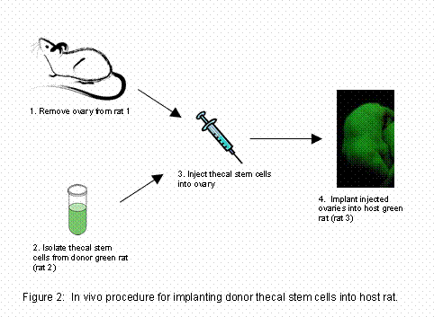
The final stage in the authors’ study was to perform intraovarian
transplantation of the thecal stem cells. The thecal cells that were used were
from “green mice” and were transplanted into the ovaries of nontransgenic mice.
Therefore the authors were able to determine if the thecal stem cells had
proliferated in the ovaries by using fluorescence microscopy. They performed
the transplantation by first removing the host ovary, injecting it with the
donor thecal stem cells and then inserting the ovaries into the empty bursa of
another host nontransgenic mouse. This procedure was replicated twice. The
reason the donor cells were not directly injected into the ovaries while still
in the animal is because this caused heavy bleeding and the donor cells did not
survive. Finally the host mice were given equine chorionic gonadotropin to
produce follicular development. When the ovaries were removed and observed the
green fluorescent cells had proliferated and surrounded the fully developed
follicles.
I found that this article informative and well written. More importantly, the
authors clearly outlined their thought processes. They began with the studies
with the intent to do something completely different and when they hit the
obstacle of the cells being somatic cells rather than germ cells. The authors
instead, used this obstacle as an opportunity to study something new. Also they
raised questions throughout the article and then performed experiments to
address their questions. For example if these were not germ cells what kind of
stem cells were they? They used microscopy and gene expression profiling. One
question is when they were applying the cells to different growth factors did
true differentiation occur or was it an artifact of the media? I believe that
these cells were actually stem cells because they were able to culture multiple
passages and showed success in their transplantation. I believe that the
addition of different growth factors created a microenvironment that facilitated
differentiation. For example in the last stage they showed that granulosa cells
were needed for final differentiation. In ovaries, granulosa cells, which are
located next to the thecal cells may therefore be cellular component of the
microenvironment for the differentiation of natural thecal cells. It would be
wonderful to see future studies in animal models for translational studies with
thecal stem cells. Also they could use these experiments to further study the
effect of the microenvironment on stem cells and perhaps apply the findings to
other types of adult stem cells.


Back |
