Back
Reference: Jae-Sung Bae, et al, BM-MSC Promote Neuronal
Networks with Functional Synaptic Transmission After Transplantation into Mice
with Neurodegeneration. Stem Cells 2007;24:1307-16.
Summarized by: Rachid Hamid and Jessica Thomas, Fall
2007
LAY SUMMARY
Bone marrow derived mesenchymal stem cells (BM-MSCs) are stem cells found in
the bone marrow and have the potential of differentiation into various cell
types provided with optimal conditions. Differentiation is a process by which
stem cells are naturally or experimentally transformed into other cell types
that possess specialized function such as neural cells that are found in the
nervous system. It has been hypothesized that MSCs cells would be able to
differentiate and give rise to neural cells within the brain (1). Researchers
believe that by transplanting these cells into damaged tissues may act as an
innovative treatment by reversing the damage. One such neurodegenerative
disorder is Niemann-Pick Disease type C (NP-C). NP is an inherited metabolic
disorder characterized by accumulation of lipids in the brain due to a defect in
cholesterol transport between brain cells (2).
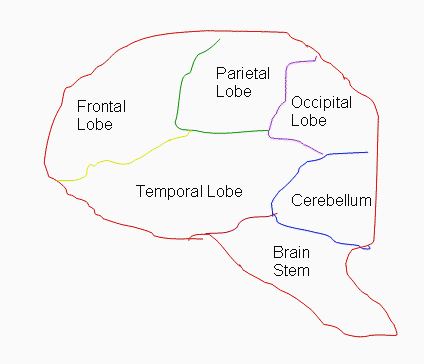
In this particular study, the scientists isolated MSCs from the bone legs of
NP-C mice and transplanted them into different groups of NP-C mice in the
cerebellum which is a region in the brain associated with motor output and
sensory perception (see brain cartoon). They also used 3T3 cells as a control
which was also transplanted into a different group of mice along with a sham
transplanted group. The use of these various groups of transplanted animals
will allow comparing and contrasting the outcomes of the experimental work and
their significance. Several experimental tools were used to analyze the samples
to prove the authors hypothesis. The authors were able to demonstrate that
transplanting MSC into NP-C mice provided significant changes at the molecular
and functional levels in one of the groups that received the MSC cells as
compared to the other groups. These changes are highlighted by the improved
balance and coordination observed in those animals which is a significant
accomplishment.
Unfortunately the results of this study concluded that
the fusion between MSCs and damaged neurons rather than, hoped for, cellular
differentiation was behind the improved symptoms of some of the mice. Although
the findings were interesting, they would have been more convincing if minor
details were not overlooked. First, one of the risks working with 3T3 cells is
the potential of uncontrollable growth and the possibility of undesirable
cancerous activity. Second, the article lacked sufficient evidence of MSCs
identity. It is crucial to isolate and identify the proper cell of interest for
subsequent use in transplant studies. Third, the authors failed to address the
long term stability and function of fused MSC. The mice received transplants at
3 weeks of age and were sacrificed 2-4 weeks later. This is far short from the
life expectance of laboratory mice of up to two years where the possibility of
side effects might appear.
http://en.wikipedia.org/wiki/Mesenchymal_stem_cell
http://www.ninds.nih.gov/disorders/niemann/niemann.htm
SCIENTIFIC SUMMARY
The goal of this summary is to present the authors’ evaluation of the
therapeutic potential of bone-marrow-derived mesenchymal cells (BM-MSC) in
neurodegenerative disease, using the Niemann-Pick type C mouse (NP-C) model. NP
is an inherited metabolic disorder characterized by the accumulation of lipids
in the brain, due to a defect in cholesterol transport among brain cells (1).
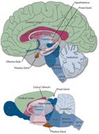
The flow chart summarizes the methods and techniques uses in the study.
 MSCs were
isolated from tibias and femurs of NP-C mice heterozygous for DsRed/Phgdh and
characterized for cluster differentiation CD29/CD90+ and for CD34/CD45/CD117-.
At 3 weeks of age BM-MSCs were transplanted into the cerebellums {see brain
cartoon} (2) of NP-C mice and NP-C GsbsGFP+. 3T3 fibroblasts cells and
sham transplanted animals were used as assay controls. MSCs were
isolated from tibias and femurs of NP-C mice heterozygous for DsRed/Phgdh and
characterized for cluster differentiation CD29/CD90+ and for CD34/CD45/CD117-.
At 3 weeks of age BM-MSCs were transplanted into the cerebellums {see brain
cartoon} (2) of NP-C mice and NP-C GsbsGFP+. 3T3 fibroblasts cells and
sham transplanted animals were used as assay controls.
Microarray analysis showed a differential gene expression
among the test groups at 2 and 4 week time points post transplantation.
Subsequent evaluation of genes of interest using quantitative real time
polymerase chain reaction (qPCR) proved increased expression of
alpha-amino-3-hydroxy-5-methyl-4-isoxazolepropionic acid (AMPA) receptors,
Gamma-aminobutyric acid (GABA) receptors, myelin and glutamate decarboxilase.
All of which have been associated with neurotrasmission in the cerebellum. MSC
transplanted animals showed increased numbers of purkinje neurons in their
cerebellums and improved Rota-Rod test outcomes for balance and coordination and
deceased lipid accumulation as compared to 3T3 and sham transplanted animals.
Transplanted animals showed slight decrease in brains size as compared to normal
animals. However, this decrease was insignificant. The electrophysiology
evaluation, the most interesting finding in this study, the authors were
succesful in proving that GFP+/DsRed+ (from NP-C GFP+ mice transplanted with
BM-MSC DsRed+ cells) purkinje cells respond to depolarizing current pulses.
These findings contrast the results of DsRed positive cells. This indicated
that BM-MSC undergone fussion with purkinje neurons rather than
transdifferentiation.
Although the author’s findings were interesting, they
would have been more convincing if minor details were not overlooked as stated
below.
The author’s characterization of BM-MSC was
insufficient by including only CD29/CD90 as positive and CD34/CD45/CD117 as
negative markers fro selection. It would be more credible and interesting if
the isolated mouse BM-MSC expressed Oct-4 and Rex-1 (3) or flowcytometry
supporting data was done and included.
3T3 are immortalized fibroblasts cells originally isolated from mouse
embryos might not be the most ideal control to use. The authors could have
differentiated the BM-MSC invitro into fibroblasts and used in the
transplant instead of 3T3. This will serve two purposes. First, showing that
the BM-MSCs are true stem cells as an additional proof. Second, the use of
BM-MSC derived fibroblasts would allow for a meaningful comparison to
BM-MSC.
The authors failed to address the long term stability and function of fusion
by BM-MSC with neural cells. The mice received transplants at 3 weeks of age
and were sacrificed 2-4 weeks later. Far short from the life expectance of
laboratory mice of up to two years.
References
- www.ninds.nih.gov/disorders/niemann/niemann.htm
- www.learn.genetics.utah.edu/.../neurobiol.cfm
- Lamoury FMJ, Croitoru-Lamoury J, Brew BJ. Undifferentiated mouse mesenchymal
stem cells spontaneously express neural and stem cell markers Oct-4 and Rex-1. Cytotherapy. 2006; 8:228-42.
Back |