Slides 14
[Lab 2] [Slides 71] [Slides 14] [Slide 62] [Slides 24] [Slides 31]

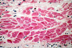
Slides 14:  Heart:  Myocardial Infarction (Recent)

path2_14l_1.jpg
path2_14l_1.jpg

A. Heart, cross section, gross.  Large area of pallor in the antero-septal region of the left ventricle represents a recent myocardial infarction.

B. Recent myocardial infarction , cross section of myocardial cells .  High power.  Note absence of myocardial cell nuclei, and edema and fragmented  (neutrophilic) debris in the interstitium

path2_14l_1.jpg

C. Recent myocardial infarction , longitudinal section of myocardial cells .  High power.  Note absence of myocardial cell nuclei and presence of polys in the interstitium.

path2_14l_1.jpg

D. Recent myocardial infarction , longitudinal section of myocardial cells .  Medium High power.  Again, note the absence of myocardial cell nuclei  in the central necrotic area and numerous polys in the interstitium.  Myocardial nuclei are seen in the viable area at the edge of the slide

On to the Slides

[Lab 2] [Slides 71] [Slides 14] [Slide 62] [Slides 24] [Slides 31]

Pathology Course Menu | Pathology | NJMed P.C. | NJMS | Search | UMDNJ

Please direct questions and comments to Alexander G. Izaguirre  izaguial@umdnj.edu