Microscopy Systems
Reserve equipment or request services now on iLabs!
Leica Stellaris 8 tau-STED Super Resolution Microscope
| Session |
Internal Price |
External Price |
| Self Use |
$35.00/hr |
N/A |
| Assisted Use/Training |
$75.00/hr |
$175.00/hr |
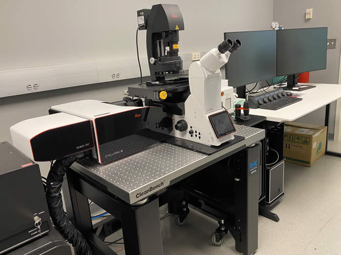
The Leica Stellaris 8 Stimulated Emission Depletion Super-Resolution microscopy system is able to achieve resolutions less than 50 nm in the lateral direction and 130 nm in the axial direction. The Stellaris 8 scan head is equipped with both standard and resonant galavonometer pairs, Leica’s aousto-optical beam splitter, tunable white light laser, with a range from 440-790 nm, and Power HyD hybrid detectors. This combination not only provides for the most versatile confocal system on the market but also enables fluorescent lifetime imaging capabilities. The system also includes two pulsed STED lasers at 775 nm for STED imaging in red and far-red channels and 589 nm for STED Imaging in the green channels. The system includes all of Leica’s STED objectives, including HC PL APO 86x/1.20 W motCORR STED W, Obj. HC PL APO 93x/1.30 GLYC motCORR STED W, 100x/1.40 Oil STED W. The system also includes Leica’s tau STED which applies the FLIM capabilities of the Stellaris 8 with STED imaging resulting in higher resolutions at low depletion laser powers. Leica claims that this system can reach resolution less than 50 nm.
If you are planning on publishing image data captured using the Leica Stellaris 8 tau-STED system, please acknowledge the National Institutes of Health High End Instrumentation Grant S10OD25182.
Nikon A1R+ HD Confocal Microscope
| Session |
Internal Price |
External Price |
| Self Use |
$30.00/hr |
$50.00/hr |
| Assisted Use/Training |
$70.00/hr |
$110.00/hr |
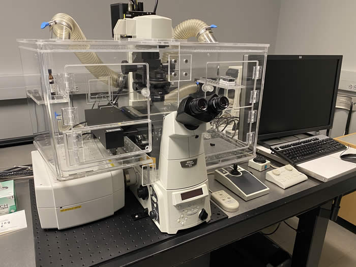
The Nikon A1R confocal microscope, has four independent channels and spectral imaging capabilities. The A1R is installed on a fully motorized Nikon Eclipse Ti inverted microscope equipped with a gas and temperature controlled environmental chamber and a perfect focus system to assist in studies involving live cultured cells. The Nikon A1R can excite a wide range of fluorochromes across the visible spectrum using the 405, 457, 488, 514, 561 and 640nm laser lines. The microscope is also equipped with filter sets for the detection of the most commonly used fluorochromes and fluorescent proteins and researchers can also make use of the spectral detector to detect any visible light emission spectra. The microscope has a wide selection of objectives for most applications including: 10x/0.45 Plan Apo, 20x/0.75 Plan Apo VC, 40x/1.0 Plan Apo Oil, 60x/1.40 Plan Apo Oil, 40x/1.25 Lambda S Water, 60x/1.27 Plan Apo VC Water, 25X/1.05 Plan Apo Silicone, 100X/1.35 Plan Apo Lambda S Silicone. The instrument was recently upgraded with Nikon’s high sensitivity GaAsP detectors and high speed high definition resonant scanner. The Nikon A1R Confocal is controlled with NIS Elements High Content Analysis Software combining the automated control of image acquisition with advanced image analysis to enable users to capture imaging data sets in significantly reduced time frames.
Nikon Ti2 HCA Inverted Fluorescence Microscope
| Session |
Internal Price |
External Price |
| Self Use |
$18.00/hr |
$28.00/hr |
| Assisted Use/Training |
$58.00/hr |
$90.00/hr |
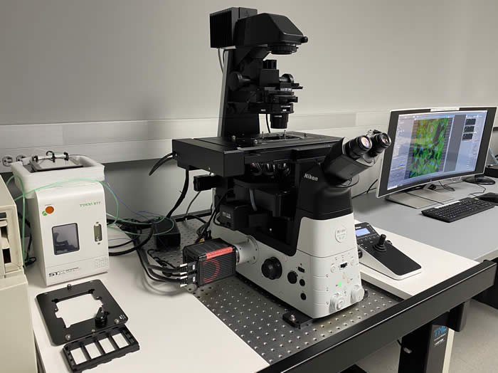
The Nikon Ti2 High Content Acquisition and Analysis System is built upon a Nikon Ti2-E fully motorized inverted research microscope including Nikon’s perfect focus system. This automation allows for the programmed automated acquisition of a variety of specimen preparations including slides, culture dishes, chambered coverglass and well plates. The encoded stage and perfect focus system in conjunction with stage top incubation will allow for the capture of multipoint extended time-lapse imaging experiments. And in addition the system is capable of the scanning of whole slides using both brightfield and fluorescence techniques. This system is also fitted with state of the art led illumination from Lumencor in both brightfield and fluorescence modes. These light sources can be trigged independently using high speed electronic pulses allowing for the tight synchronization of the light source to the camera leading to extremely high acquisition rates. Tying these hardware pieces together is the unique Nikon NIS Elements High Content Analysis software which combines advanced image analysis techniques with programmed image acquisition resulting in the combined automation of both image capture and analysis resulting in higher quality analyzed data captured in significantly reduced time frames.
Zeizz Axioscan7
| Slide |
Internal Price |
External Price |
| Brightfield |
$2.50/slide |
$5.75/slide |
| Flourescent - 1 Channel |
$2.50/slide |
$5.75/slide |
| Flourescent - 2 Channel |
$5.00/slide |
$11.50/slide |
| Flourescent - 3 Channel |
$7.50/slide |
17.25/slide |
| Flourescent - 4 Channel |
$10.00/hr |
$23.00/slide |
| Z-Stack |
$1.00/additional focal plane |
$1.50/additional focal plane |
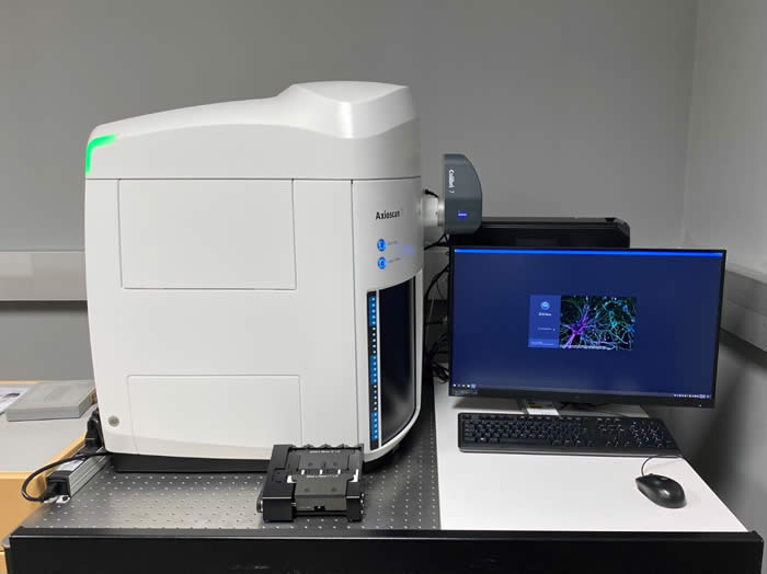
The Zeiss Axioscan 7 is a fully automated slide scanning microscopy system able to capture full slide scans of both standard brightfield illuminated histology slides as well as slides labeled with multiple fluorescent dyes. The instrument is equipped with two objective lenses a 5x Fluar NA 0.25 for low resolution scans to aid in the identification of regions of interest and a 20x Plan Apochromat NA 0.8 for the high-resolution scanning of the tissue sections. The instrument is equipped with a five-megapixel Zeiss Axiocam 705 color camera for brightfield imaging and a 12-megapixel Zeiss Axiocam 712 monochorome camera for fluorescent imaging. For fluorescence illumination, the Axioscan 7 is equipped with the Zeiss Colibri 7 solid state light source. The Colibri 7 is equipped with 5 independent LED lamps for the excitation of DAPI, FITC, TRITC, Texas Red, CY5 or spectrally similar dyes. The intensity of each LED lamp can be independently controlled through software ensuring optimized exposure and the reduction of photobleaching. In addition, the LEDs can be rapidly triggered to enable the high-speed capture of multichannel images reducing the time required to capture large tiled multichannel images. The Axioscan 7 system can automatically detect regions on a slide containing tissue sections and only scan those regions of interest saving instrument time and data storage. The system can automatically create focus surface maps and then adjust focus during scanning to ensure that all regions of tissue section are in focus even if the sample itself is uneven.
Keyence BZ-X710 Inverted Fluorencence Microscope
| Session |
Internal Price |
External Price |
| Self Use |
$12.00/hr |
$20.00/hr |
| Assisted Use/Training |
$52.00/hr |
$82.00/hr |
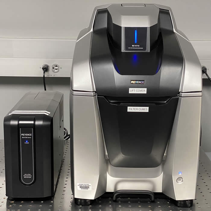
The Keyence BZ-X710 All-in-One Fluorescent Microscope is a fully motorized inverted microscope capable of capturing fluorescence, brightfield and phase contrast images of biological specimens on a variety of media including, microscope slides and culture dishes and plates. The motorization and intuitive software interface enable the rapid acquisition of complex microscopic imaging experiments including multichannel fluorescence, high resolution tiled images and the high-throughput capture of multi-well plates. All of this is achieved in a compact integrated microscopy system that is at home on the lab bench, requiring no specialized darkroom.
Nikon Eclips 50i Brightfield Microscope
| Session |
Internal Price |
External Price |
| Self Use |
$10.00/hr |
$16.00/hr |
| Assisted Use/Training |
$50.00/hr |
$78.00/hr |
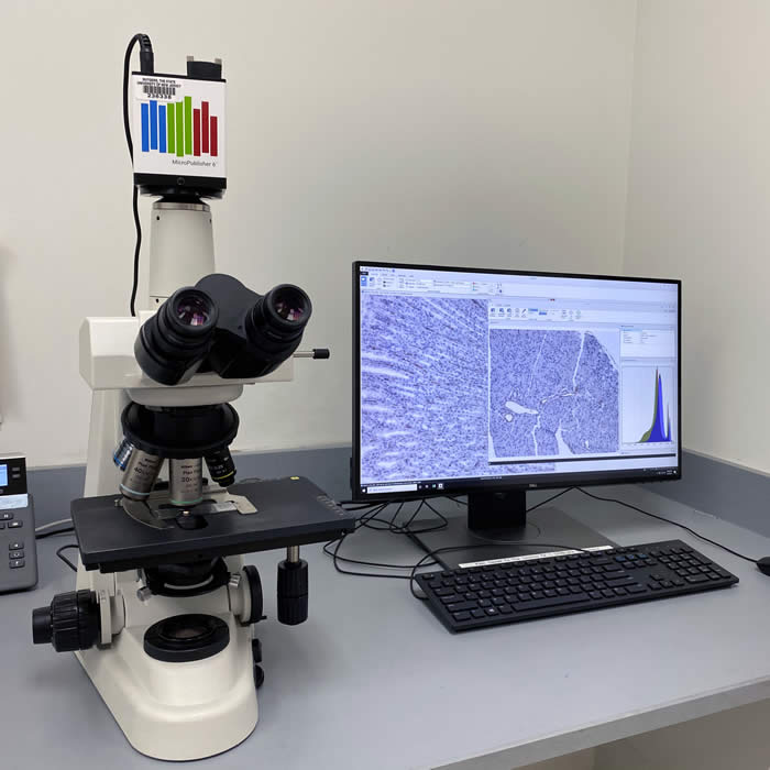
The Nikon Eclipse 50i is routine brightfield microscope for the documentary capture of histological slides. It is equipped with 4x, 10x, 20x, 40x and 60x objective lenses and the Q imaging Micropublisher 6 five MP USB 3.0 digital camera. The computer controlling the camera is also equipped with Media Cybernetics Image Pro Image Analysis software platform.
Objective Imaging Slide Scanning System
| Session |
Internal Price |
External Price |
| Self Use |
$10.00/hr |
$16.00/hr |
| Assisted Use/Training |
$50.00/hr |
$78.00/hr |
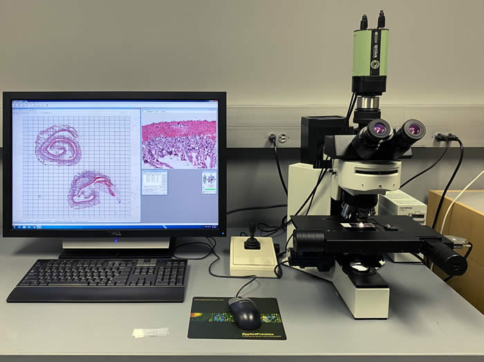
The Objective Imaging slide scanning system is for the capture of large brightfield tiled whole slide images. The system is installed on an Olympus BX upright microscope with a 2x and 20x lenses for preview and high-resolution images respectively. The system uses the Objective Imaging OASIS PCI card and Prior motorized stage to capture whole slide images with short acquisition times.
Reserve equipment or request services now on iLabs!