|
|
 |
|
|
23. BREAST: CARCINOMA
PERTINENT CHANGES:
In some areas a normal portion of breast is present and is composed of interlobular and terminal ducts.
Most of the tissue is occupied by epithelial tumor cells arranged in cords and sheets, sometimes surrounding intact ducts.
The nuclei of the tumor cells vary in size, shape and staining character. The nucleocytoplasmic ratio is altered in favor of the nuclei.
Pink granular zones of necrosis are present; one area involves a duct.
Irregular bands of connective tissue lie among the tumor cells (desmoplasia).
The carcinoma extends irregularly into surrounding fat and also muscles. |
|
|
|
23A. "See Robbins, Pathologic Basis of Disease, Cotran, Kumar, and Collins, 6th edition; Figure 25-16a, p. 1108." |
|
|
|
|
|
|
|
|
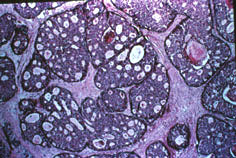
23B. Low power showing cribriform intraductal growth of ductal cells with central areas of necrosis |
|
|
|
23C. High power shows the cells comprising the cribriform intraductal growth are monomorphic ductal cells (no myoepithelial cells) |
|
|
23D. Invasive ductal carcinoma, medium power. Another area of the tumor shows invasive ductal carcinoma with irregular ducts lined by hyperchromatic cells infiltrating a fibrotic stroma |
|
|
|
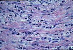
23E. High power of infiltrating lobular carcinoma. Relatively small cells singly or in single file infiltrate fibrotic stroma. Compare with infiltrating ductal carcinoma |
|
|
|