|
|
 |
|
|
43. BREAST: SCLEROSING ADENOSIS (FIBROCYSTIC CHANGES)
PERTINENT CHANGES:
There is a diffuse fibrosis which compresses and distorts an overgrowth of lobular epithelial cells.
Many of the ductules are dilated and filled with a light blue material.
The ductules show an increase in the number of cells.
There are cystically dilated ducts filled with a blue fluid.
There are also dilated glands lined by apocrine cells. |
|
|
|
|
|
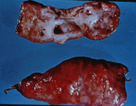
43A. Gross specimen of fibrocystic change with numerous, variously sized, smooth walled cysts in fibrotic breast tissue |
|
|
|
|
43B. "See Pathology, Rubin and Farber, 2nd edition; Figure 19-5, p. 980." |
|
|
|
|
|
|
|
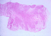
43C. Scanning power of sclerosing adenosis. Lobular pattern of ductules (acini) is maintained in dense fibrous tissue |
|
|
|
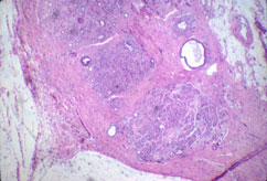
43D. Low power. Increased numbers of acini comprise the lobules, which show perilobular and intralobular fibrosis |
|
|
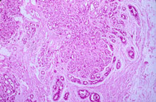
43E. Medium power. Although distorted by the fibrous tissue, lobular pattern is preserved. Central acini are compressed while peripheral ones are slightly dilated |
|
|
|