This was developed by Alexander G. Izaguirre & Dr. Anthony Grygotis

 Slides 91
[Lab 6] [Slides 73] [Slides 25] [Slides 66] [Slides 28] [Slides 16] [Slides 57] [Slides 91] [Slides 43] [Slides 23]

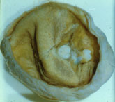
91.OVARY:  DERMOID CYST

PERTINENT CHANGES:

  1. A narrow portion of the cortex of the ovary is present in some of the slides.
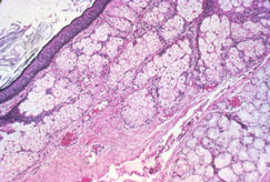
  2. The majority of the structure is composed of epidermis and dermis with skin adnexa among which are sebaceous glands, sweat glands and hair shafts.
  3. In some slides inclusion cysts are present.  These are lined by squamous epithelium.  The lumens contain keratin.
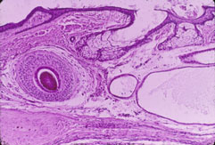
  4. Some slides contain structures resembling a bronchus lined by tall columnar epithelium.  A portion of well-developed cartilage lies in the wall and in addition, the wall contains muscle fibers.  The lumen contains pink-blue amorphous material.
  5. Nerve structures are present in some of the slides.  Bone is present in some slides.
  6. Note brown pigment in basal layer of epidermis and in the superficial dermis

91A.  Gross photograph of opened dermoid cyst (benign cystic teratoma) of the ovary.  Cyst has a tan lining (see next images)and contains several identifiable teeth

91B.  Low power of cyst lining which is mainly skin consisting of stratified squamous epithelium representing epidermis with sebaceous glands and hair follicles.  A small portion of nervous tissue is present at the lower left

91C.  Medium power has squamous epithelium and sebaceous glands with other glands resembling Brunner's glands of the duodenum.  The predominant tissue is skin, hence the name "dermoid", but representation of other tissues from all three germ layers is usually seen in these cases

Previous                                        Next

[Lab 6] [Slides 73] [Slides 25] [Slides 66] [Slides 28] [Slides 16] [Slides 57] [Slides 91] [Slides 43] [Slides 23]

Pathology Course Menu | Pathology | NJ Med. P.C. | NJMS | Search | UMDNJ

Please direct comments and questions about the Pathology Medical Student pages to izaguial@umdnj.edu Thank You