|
|
 |
|
|
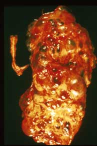
Slide 40: Polycystic Disease (Adult, Autosomal Dominant)
PERTINENT CHANGES
- The normal architecture is completely disrupted by very large cysts.
- The cysts are present in the cortex and medulla.
- They are lined by flattened cells in some regions and dense connective tissue in others.
- There is a great decrease in the number of glomeruli.
- Tubules are decreased in number and are distorted by an increase of interstitial connective tissue.
|
|
|
|
|
|
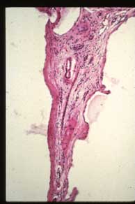
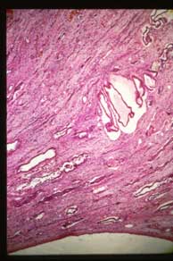
C. Low power showing cysts, present in cortex and medulla, have disrupted and replaced most normal structures and are accompanied by fibrous tissue. |
|
|
|
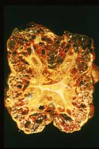
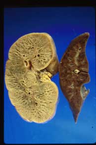
E. This image is a cut surface view of the liver and kidney from a patient with autosomal recessive (childhood) polycystic kidney disease. Numerous uniform, small cysts (elongated and at right angles to the cortical surface) are in the cortex and medulla of the kidney giving it a spongelike appearance. Multiple cysts and fibrosis occur in the liver. |
|