|
|
 |
|
|
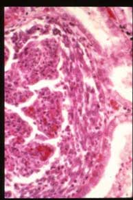
Slide 70: Glomerulonephritis, Rapidly Progressive
PERTINENT CHANGES
- The capsular surface is fairly even.
Interstitial tissue shows occasional small focal collections of lymphocytes and
mononuclear cells.
- Arteries and arterioles appear normal.
Glomeruli:
- Most glomeruli are intact and Bowman's space is patent.
- An occasional glomerulus shows a small pink granular zone of necrosis.
- Around these zones is an increase of cells (endothelial and mesangial)
- An occasional glumerulus shows focal hyaline zones of fibrosis (scar)
- Some glomeruli show proiferation of epithelial cells (cresents)
- An occasional glomerulus shows a large increase of cells with obliteration of the Bowman's space.
- Tubules:
- A light pink precipitate is present in some tubules.
- Focal collection of RBC's in some tubules.
- The cytoplasm of some proximal convoluted tubule cells is swollen and granular
|
|
|
|
Images: |
|
|
|
|
|
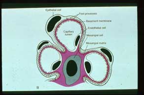
Intro. A. Diagram of ultrastructure of normal glomerulus. The basement membrane lies between epithelial and endothelial cells and is incomplete where it abuts the mesangial matrix, which lies in an intracapillary location. Note epithelial cells with discrete foot processes, endothelial cells with slit pores, and mesangial cells. |
|
|
|
|
|
|
|
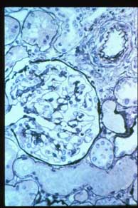
Intro B. Normal glomerulus, silver stain. Note thin even staining glomerular basement membrane of uniform thickness. |
|
|
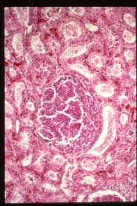
B. Medium power of a hypercellular glomerulus showing a well developed epithelial crescent. |
|
|
|
|
|
|
|
|
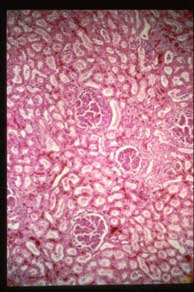
A. Low power overview of renal cortex showing generally intact hypercellular glomeruli, some of which show a crescentic proliferation of parietal epithelial cells lining Bowman's capsule (center). |
|
|
C. High power of previous image showing the epithelial crescent and some inflammatory cells in the glomerulus. |
|
|