|
|
 |
|
|
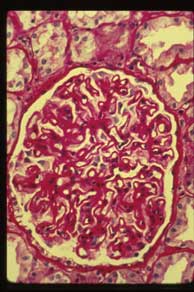
Slide 51: Membranous Glomerulopathy
PERTINENT CHANGES
- A slight depression of the cortical surface is seen.
- Interstitial tissue is widened by an increase of loose connective tissue separating renal tubules and there are focal collections of lymphocytes and mononuclear cells.
- Arterioles are moderately thickened by pink hyaline material; arteries are thickened by an increase of intimal connective tissue
- Glomeruli:
- The capsules of some glomeruli are thickened by connective tissue.
- The glomeruli are enlarged and contain variable amounts of pink material. In some there is almost complete replacement of glomeruli by the thickened basement membranes.
- Tubules: Changes are striking.
- They are of irregular size and shape.
- Occasional tubule decreased in size, others are irregularly dilated and lined with flattened cells.
- Hyaline casts in many lumens.
- Proximal convoluted tubules show swelling of cytoplasm and vacuolization. Much lipid in tubules.
|
|
|
|
|
|
|
|
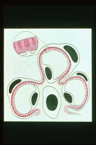
A. Diagram of renal glomerular ultrastructure with membranous glomerulopathy showing multiple, small deposits (darker red) on the epithelial side of the basement membrane (stippled red). Basement membrane material grows between and over these deposits (pink) creating the appearance of a thickened glomerular basement membrane (GBM). Note fused epithelial foot processes. Compare to first image of this unit (diagram of normal glomerulus). |
|
|
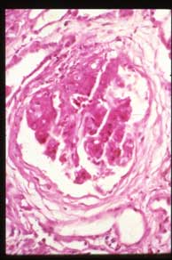
C. H&E stain, high power |
|
|
|
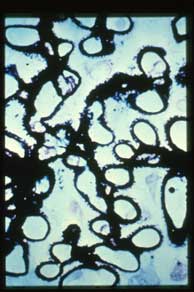
E. High power of a Jones silver stain showing the typical "spike and dome" pattern. The silver stains the basement membrane material (the heavy line and spikes) but not the immune deposits (the spaces between the spikes). The latter would stain with immunofluorescene microscopy. |
|
|
|
|
|
|
|
|
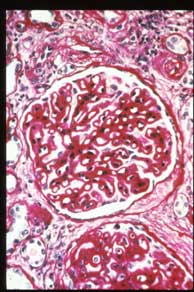
B. PAS stain, high power shows markedly thickened GBMs of membranous glomerulopathy |
|
|
D. PAS stain, high power, of another typical case of idiopathic membranous glomerulopathy |
|
|