|
|
| [Lab 3] [Slides 70] [Slides 51] [Supplement] [Slide 41] [Slides 40] [Slides 69] [Slides 87] |
|
|
||||||||||||||||||||||||||||||||||||||||
|
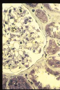
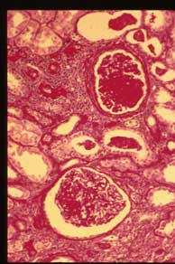
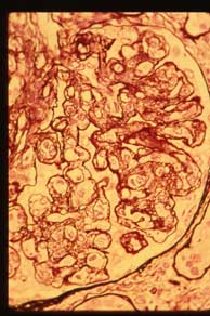
Membranoproliferative Glomerulonephritis (MPGN) |
|
|
|
| [Lab 3] [Slides 70] [Slides 51] [Supplement] [Slide 41] [Slides 40] [Slides 69] [Slides 87] |
|
Pathology Course Menu | Pathology | NJ Med, P.C. | NJMS | Search | UMDNJ |
|
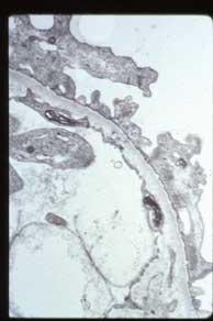
G. Electron micrograph of glomerulus. Ultrastructural examination reveals fusion of epithelial foot processes but no other abnormalities. |
|||
|
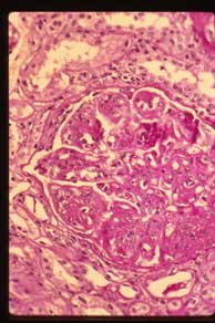
J. Jones silver stain shows "splitting" of the GBM brought about by subendothelial interposition of mesangial cells and deposition of new basement membrane material beneath the endothelium, giving rise to the "tram track" appearance. |
|||