Slides 69
[Lab 3] [Slides 70] [Slides 51] [Supplement] [Slide 41] [Slides 40] [Slides 69] [Slides 87]

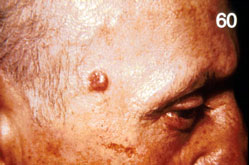
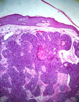
69.  SKIN:  BASAL CELL CARCINOMA

PERTINENT CHANGES:

  1. Lying in superficial corium are nests of closely packed cells.
  2. They are uniform in size with oval and dark nuclei and are surrounded by a rim of similar cells which resemble the lower-most layer of epidermis and hair follicles.
  3. These foci are present in different parts of the corium; they are separated from the overlying epidermis.
  4. Mitotic figures are present.


slide_69_1.jpg
slide_69_2.jpg

A. Clinical picture of a basal cell carcinoma of the upper face presenting as a typical pearly, telangiectatic nodule.

B. The tumor is composed of nests of basaloid cells of uniform size in the dermis.

Previous                                   Next

[Lab 3] [Slides 70] [Slides 51] [Supplement] [Slide 41] [Slides 40] [Slides 69] [Slides 87]

Pathology Course Menu | Pathology | NJ Med, P.C. | NJMS | Search | UMDNJ

Please direct comments and questions about the Pathology Medical Student pages to izaguial@umdnj.edu.  Thank you.