Slides 87
[Lab 3] [Slides 70] [Slides 51] [Supplement] [Slide 41] [Slides 40] [Slides 69] [Slides 87]

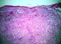
87.  SKIN:  MALIGNANT MELANOMA

PERTINENT CHANGES:

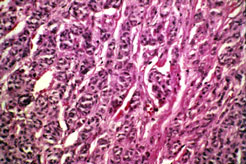
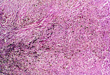
  1. Large masses of tumor cells replace and distort normal architecture.
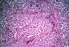
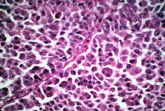
  2. These cells
    1. Are heavily pigmented in some areas, much less so in others.
    2. Have a tendency to occur in small thecae.
    3. Adhere to each other poorly, if at all.
    4. Have large, heterogeneous, hyperchromatic atypical nuclei with prominent magenta nucleoli.
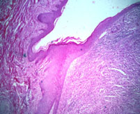
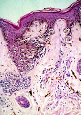
  3. Similar appearing cells are occasionally present in junctional nests and in the stratum basalis of the epidermis overlying the tumor.
  4. There is no significant lymphocytic infiltrate associated with this tumor.
  5. In places, a thickened stratum malpighii and granulosum and markedly thickened and condensed stratum corneum identify this as volar skin.
slide_87_1.jpg
slide_87_2.jpg
slide_87_3.jpg
slide_87_4.jpg

A. Low power photomicrograph showing nests of tumor cells extending from an ulcerated epidermis in nodular aggregates (vertical growth phase).

B. Edge of the tumor shows it to project above the skin surface as well as invading into the dermis.

C.  Medium power.  Tumor cells are poorly cohesive.

D.  Higher power.  Nuclei are large, hyperchromatic, and contain prominent nucleoli. 

slide_87_5.jpg

E.  In some areas, tumor cells form small aggregates called theques.

slide_87_6.jpg

F.  Melanin pigmentation is seen in some portions of the tumor.

slide_87_7.jpg

G.  Shown are tumor cells in the dermal epidermal junction region which illustrates the radial growth phase, which may precede the vertical growth phase   

Previous

[Lab 3] [Slides 70] [Slides 51] [Supplement] [Slide 41] [Slides 40] [Slides 69] [Slides 87]

Pathology Course Menu | Pathology | NJ Med, P.C. | NJMS | Search | UMDNJ

Please direct comments and questions about the Pathology Medical Student pages to izaguial@umdnj.edu.  Thank you.