Cardiovascular Imaging Fellowship
Facilities and Resources

University Hospital, the principal teaching hospital of Rutgers New Jersey Medical School (NJMS), has 519 licensed inpatient beds and a full service echocardiography lab, nuclear cardiology lab, cardiac computed tomography, and cardiac magnetic resonance imaging. All the cardiac imaging services at University Hospital are staffed with fulltime Rutgers NJMS faculty who have advanced training and expertise and board certifications in these areas.
Echocardiography:
The laboratory includes an inpatient and outpatient facility. The fellow will develop expertise in all areas of transthoracic echocardiography, including 2D, M-mode, color and spectral Doppler. This includes contrast imaging, harmonic imaging, tissue Doppler, strain imaging and three-dimensional (3D) imaging. The fellow will also develop expertise in performing and evaluating tranesophageal (TEE) studies with a clinical focus on native valve disease, including evaluation for potential transcatheter interventions; prosthetic valve function/dysfunction; infective endocarditis and its complications; cardiac source of embolus; and intraprocedural TEE imaging for structural heart cases.
The fellow will also become proficient in performing and reporting exercise and pharmacological stress echocardiography including for coronary artery disease / ischemia, preoperative assessment, and valve disease evaluation.
Nuclear Cardiology:
The laboratory is fully integrated within the Nuclear Medicine division with a dedicated treadmill and hot lab for handling radiopharmaceutical doses. The fellow will develop expertise in all areas of nuclear cardiology, including planar, SPECT, PYP, viability testing, and FDG PET for viability, cardiac sarcoidosis, and inflammation / infection.
Cardiac CT:
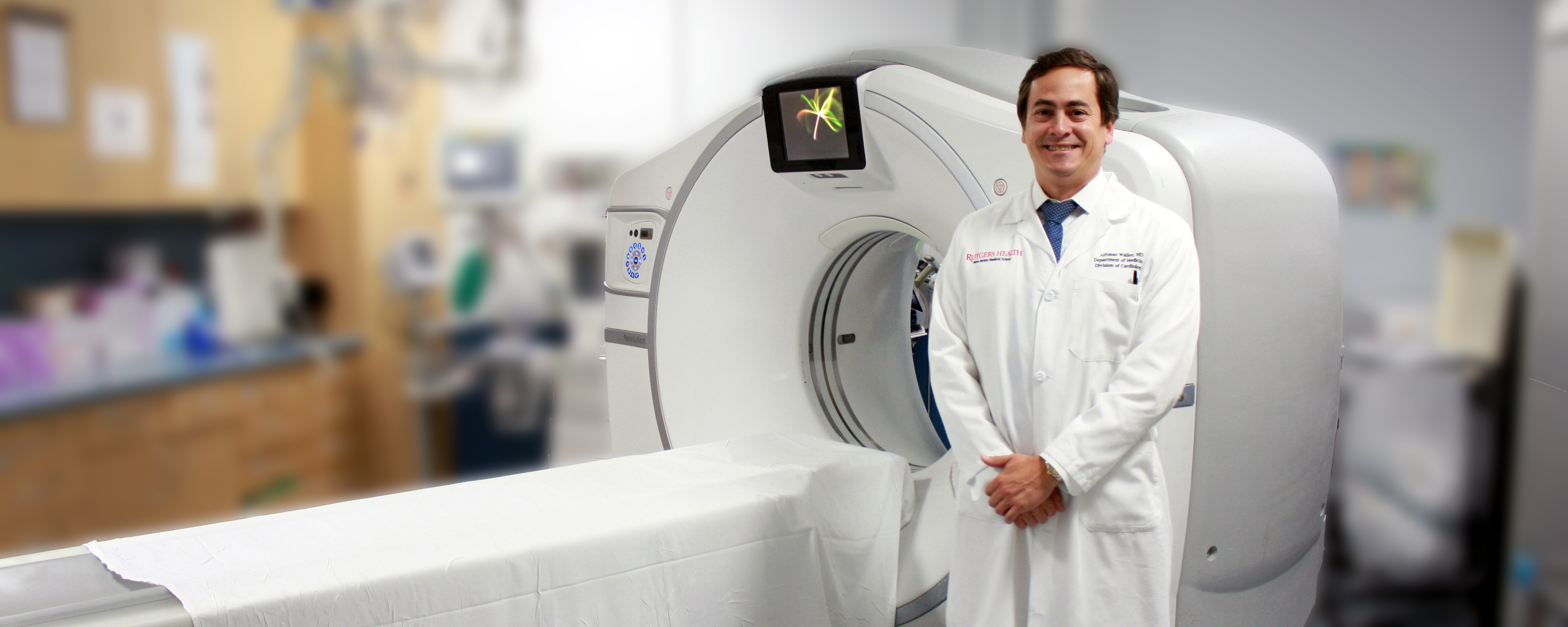
The fellow will develop expertise in all areas of cardiovascular CT acquisition, supervision, and interpretation, including laboratory requirements, radiation safety, contrast administration, pharmacologic interventions, and utilizing a 3D workstation. The lab performs CTs for coronary calcification, coronary arteries, pulmonary vein evaluation, left atrial appendage evaluation, tumors and masses of the heart, pericardial disease, and valvular heart disease, including pre-TAVR evaluation.
Cardiac MRI:
The fellow will develop expertise in all areas of cardiovascular MRI acquisition, supervision, and interpretation, including laboratory requirements, contrast administration, pharmacologic interventions, and utilizing a 3D workstation. The lab performs cardiac MRIs quantitative assessment of left and right ventricular size and function, myocardial perfusion, myocardial disease and viability, pulmonary vein evaluation, tumors and masses of the heart, pericardial disease, and valvular heart disease.

