The Healthy Heart
The Healthy Heart
Anatomy of the Heart
The heart is a muscle located in the middle of the chest and behind the breastbone (sternum) that is approximately the size of a fist. It is "powered" by an "electrical system" that signals the heart muscle to beat rhythmically approximately 72 times per minute.
If you were to slice it down the middle, you would find that it has three layers, the endocardium (the smooth inside lining of the heart); the myocardium (the muscle layer of the heart); and the epicardium (the outside lining of the heart). The pericardium is the tough, fluid-filled sac that surrounds the heart itself, this pseudo-fourth layer provides protection and minimizes the friction created by the heart beat.
The Heart is divided into four chambers: The Right Atrium (RA), the Right Ventricle (RV), the Left Atrium(LA) and the Left Ventricle (LV). The electraical signal for each heartbeat begins in the Right Atrium in an area called the sinus node (aka the heart's natural pacemaker). Each chamber has a one-way valve at its exit that prevents blood from flowing backwards. When each chamber receives an electrical pulse, it contracts, and the valve at its exit opens pumping blood through it and when it is finished contracting the valve closes. As the lower chambers fill with blood, the electrical signal travels along special conduction tissues to the AV node, where it pauses for a few seconds, allowing the chambers to finish filling.
There are four valves in the heart including the Tricuspid Valve, which is at the exit of the Right Atrium, the Pulmonary Valve, which is at the exit of the Right Ventricle, the Mitral Valve, which is at the exit of the Left Atrium and the Aortic Valve, which is at the exit of the Left Ventricle.
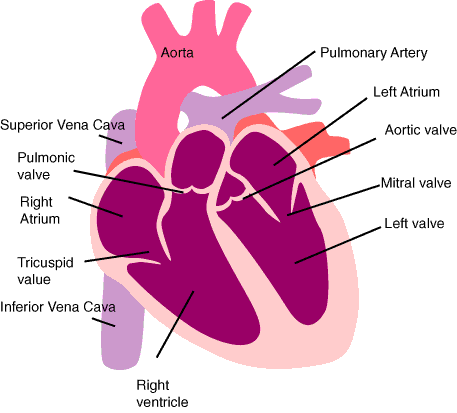
When the heart muscle contracts (or beats) it pumps blood out of the lower chambers of the heart. The heart contracts in two stages. In the first stage the Right and Left Atria contract at the same time, pumping blood to the Right and Left Ventricles. Then the Ventricles contract together (called systole) to propel blood out of the heart. After this second stage, the heart muscle relaxes (called diastole) before the next heartbeat. During this time, the muscle resets itself for contraction and blood fills the atria.
Functions of the Heart
The right side of the heart collects oxygen-poor blood from the body and pumps it to the lungs where it picks up oxygen and releases carbon dioxide while the left side collects oxygen rich blood from the lungs and pumps it to the body so that the cells throughout your body have the oxygen they need to function properly.
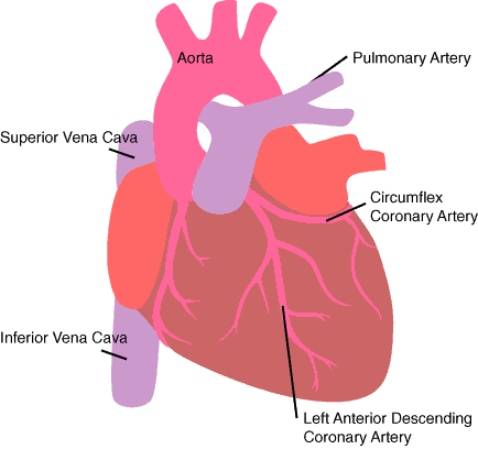
All blood enters the right side of the heart through two veins, the Superior Vena Cava(SVC), which collects blood from the upper half of the body and the Inferior Vena Cava (IVC), which collects blood from the lower half of the body.
When the heart is pumping, blood flows from the body to the Superior and Inferior Vena Cava then to the Right Atrium through the Tricuspid Valve. It flows to the Right Ventricle through the Pulmonary Valve through the Pulmonary Artery to the Lungs.
There, the blood picks up oxygen and drops off carbon dioxide in the lungs, and then flows from the lungs through the Pulmonary Veins to the Left Atriumthrough Mitral Valve to the Left Ventriclethrough the Aortic Valve to the Aorta through the two main coronary arteries -- the Left Coronary Artery (which divides into two - the Left Anterior Descending Artery and the Circumflex Artery) and the Right Coronary Artery. From here blood flows the arterial system to the body.
The heart, just like any other organ, requires blood to supply it with oxygen and other nutrients so that it can do its work. The heart does not extract oxygen and other nutrients from the blood flowing inside it. The heart gets its blood from coronary arteries, located on the outside surface of the heart, that eventually carry blood within the heart muscle through a network of branches.